 |
See our home page for detailed info on causes and
treatment of autoimmune disorders
Women and
Autoimmune Diseases1
DeLisa Fairweather*
and Noel R. Rose*
*Johns Hopkins University, Baltimore, Maryland, USA
Read our e-book for SIMPLE ANTIBIOTIC treatment protocols on
AUTOIMMUNE disease syndromes.
.Contact us for help
Autoimmune diseases affect approximately 8% of the
population, 78% of whom are women. The reasons for the
high prevalence in women are unknown, but circumstantial
evidence links autoimmune diseases with preceding
infections. Animal models of autoimmune diseases have
shown that infections can induce autoimmune disease. For
example, coxsackievirus B3 (CB3) infection of
susceptible mice results in inflammation of the heart
(myocarditis) that resembles myocarditis in humans. The
same disease can be induced by injecting mice with heart
proteins mixed with adjuvant(s), which indicates that an
active infection is not necessary for the development of
autoimmune disease. We have found that CB3 triggers
autoimmune disease in susceptible mice by stimulating
elevated levels of proinflammatory cytokines from mast
cells during the innate immune response. Sex hormones
may further amplify this hyperimmune response to
infection in susceptible persons, which leads to an
increased prevalence of autoimmune diseases in women.
Autoimmune diseases are the third most common category of
disease in the United States after cancer and heart disease;
they affect approximately 5%–8% of the population or 14–22
million persons . Autoimmune diseases
can affect virtually every site in the body, including the
endocrine system, connective tissue, gastrointestinal tract,
heart, skin, and kidneys. At least 15 diseases are known to
be the direct result of an autoimmune response, while
circumstantial evidence implicates >80 conditions with
autoimmunity . In several instances,
such as rheumatoid arthritis, multiple sclerosis, and myocarditis,
the autoimmune disease can be induced experimentally by
administering self-antigen in the presence of adjuvant
(collagen, myelin basic protein, and cardiac myosin,
respectively) . An important unifying theme in autoimmune
diseases is a high prevalence in women . Conservative
estimates indicate that 6.7 million or 78.8% of the persons
with autoimmune diseases are women .
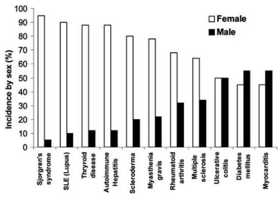
Soon after autoimmune diseases were first recognized more
than a century ago, researchers began to associate them with
viral and bacterial infections. Autoimmune diseases tend to
cluster in families and in individuals (a person with one
autoimmune disease is more likely to get another), which
indicates that common mechanisms are involved in disease
susceptibility. Studies of the prevalence of autoimmune
disease in monozygotic twins show that genetic as well as
environmental factors (such as infection) are necessary for
the disease to develop. Genetic factors
are important in the development of autoimmune disease,
since such diseases develop in certain strains of mice
(e.g., systemic lupus erythematosus or lupus in MRL mice)
without any apparent infectious environmental trigger.
However, a body of circumstantial evidence links diabetes,
multiple sclerosis, myocarditis, and many other autoimmune
diseases with preceding infections . More often, many different
microorganisms have been associated with a single autoimmune
disease, which indicates that more than one infectious agent
can induce the same disease through similar mechanisms . Since infections generally occur well
before the onset of symptoms of autoimmune disease,
clinically linking a specific causative agent to a
particular autoimmune disease is difficult . This difficulty raises the question of whether
autoimmune diseases really can be attributed to infections.
Are Autoimmune
Diseases Caused by Infections?
To address the question of whether autoimmune diseases
can be induced by infections, first autoimmunity needs to be
defined. Autoimmune disease occurs when a response against a
self-antigen(s) involving T cells, B cells, or
autoantibodies induces injury systemically or against a
particular organ. Understanding of autoimmune diseases is
hindered by the fact that some level of autoimmunity, in the
form of naturally occurring autoantibodies and self-reactive
T and B cells, is present in all normal persons . Thus, on a
proportional basis, developing autoimmune disease is the
relatively uncommon consequence of a common autoimmune
response. Although an autoimmune response occurs in most
persons, clinically relevant autoimmune disease develops
only in susceptible persons .
Given those circumstances, how can infections induce
autoimmune disease? A mechanism often called on to explain
the association of infection with autoimmune disease is
"molecular mimicry," that is, antigens (or more properly
epitopes) of the microorganism closely resemble
self-antigens. The induction of an immune response to the
microbial antigen thus results in cross-reaction with
self-antigens and induction of autoimmunity .
Although epitope-specific cross-reactivity between microbes
and self-tissues has been shown in some animal models ,
molecular mimicry has not been clearly demonstrated to occur
in human diseases . Another
possibility is that microorganisms expose self-antigens to
the immune system by directly damaging tissues during an
active infection. This mechanism has been referred to as the
"bystander effect" . However,
whether pathogens mimic self-antigens, release sequestered
self-antigens, or both, is difficult to determine.
In addition to antigen-specific mechanisms, nonspecific
mechanisms could also lead to autoimmunity after infection .
Activation of the innate immune system is essential for a
protective adaptive immune response to develop; and vaccines
that lack intrinsic activation of innate immunity (e.g.,
subunit vaccines) require microbial adjuvants to be
immunogenic . Historically, adjuvants
are considered to stimulate immune responses
nonspecifically. A renewed understanding of the critical
role of innate immunity in influencing the development of an
adaptive immune response has led researchers to a better
understanding of "the adjuvant effect" .
Although innate immune cells do not respond to specific
antigenic epitopes on pathogens, they do produce restricted
responses to particular classes of pathogens through
pattern-recognition receptors (PRR), such as Toll-like
receptors (TLR) . Interaction of the
microorganism component of adjuvants with PRR on innate
immune cells results in activation of antigen-presenting
cells and upregulation of molecules essential for antigen
presentation, such as major histocompatibility complex (MHC)
class II and B7–1/2, as well as production of
proinflammatory cytokines. This activation of PRR by the
microbial components of adjuvants stimulates the immune
response in a manner similar to pathogens such as bacteria
or viruses . The pathogen-specific
innate immune response is not the same as the nonspecific
activation that occurs after mechanical tissue damage, such
as during surgery. During mechanical injury, self-antigens
and cytokines are released without consistently stimulating
pathogen-specific responses. Autoimmune disease rarely
develops and usually resolves spontaneously, as seen in postcommissurotomy syndrome (or postcardiotomy syndrome).
Adjuvants (usually bacterial, e.g., Mycobacterium in
complete Freund's adjuvant) activate the innate immune
response in the same pathogen-specific manner when
administered with self-antigen; this process results in
organ-specific autoimmune disease in animal models .
Adjuvant alone (without self-antigen) does not usually
result in autoimmune disease, and microorganisms likely
provide not only the adjuvant effect to stimulate the immune
response but also the damage necessary to make self-antigens
available to the immune system, resulting in autoimmune
disease.
To determine whether infection can lead to autoimmune
disease, direct evidence (e.g., the ability to transfer
autoimmune disease), indirect evidence (e.g., the ability to
reproduce autoimmune disease in animal models), and
circumstantial evidence (e.g., the association of
autoantibodies with disease in appropriate clinical
settings) should be considered (3,6). The
best evidence so far that infections can induce autoimmune
diseases comes from animal models. In most animal models of
autoimmunity, including myocarditis, disease has been
transferred to naïve animals with autoimmune cells
(splenocytes or T cells), autoantibodies (7),
or both, which provides compelling evidence that infections
induce autoimmune diseases by immune-mediated mechanisms.
Lessons from
Coxsackievirus B3 (CB3) Myocarditis
Genetics of
Susceptibility to Myocarditis
Genetic background accounts for only about one third
(30%–35%) of the risk of autoimmune disease
This estimate is based on studies that compared genetically
identical, monozygotic twins to nonidentical, dizygotic
pairs, for which the concurrence rate can be as low as 2% to
7% . Therefore, noninherited factors
account for the remaining (approximately 70%) risk of
developing an autoimmune disorder. Yet, even identical twins
do not have identical immune systems. Genes outside of the
MHC contribute to the risk for autoimmune disease. However,
little information is available about the function of these
non-MHC genes. Recent studies have focused on regulatory
signals, and considerable evidence exists that cytotoxic
lymphocyte antigen–4 (CTLA-4), which provides a
downregulatory signal, influences susceptibility to
autoimmunity . Genes that involve apoptosis, a common
pathway by which immune responses generally are terminated,
may also predispose persons to autoimmune disease .
To better understand the relationship between infection
and autoimmune disease, we established a mouse model of
myocarditis, or inflammation of the heart, induced by CB3
infection . CB3 is believed to account
for most cases of myocarditis in North America and Europe;
myocarditis also leads to dilated cardiomyopathy, which can
result in heart failure and the need for a heart transplant
. The same strain of CB3 that induces myocarditis in humans also induces disease in mice, which
makes it an ideal pathogen to study. We found that
susceptibility to myocarditis is due primarily to genes that
are not part of the MHC . Our initial
investigations into the genetic predisposition of autoimmune myocarditis involved infecting many inbred mouse strains
with CB3 . Genetic analysis comparing
susceptibility loci between susceptible and resistant
strains of mice found that susceptibility to myocarditis is
associated with genes on mouse chromosomes 1 and 6 that have
been previously associated with other autoimmune diseases in
mice and humans (M. Guler and N.R. Rose, unpub. data). In
susceptible strains of mice ( e.g., BALB/c, A/J), acute
heart inflammation develops approximately 7–12 days after
infection; the inflammation resolves by day 21, and then a
chronic phase of inflammation and dilated cardiomyopathy
develops from day 35 . In contrast, only
the acute phase of the disease develops in resistant strains
like C57BL/6. After mice are infected with CB3,
autoantibodies are produced against cardiac myosin, the
major component of heart muscle. We found that susceptible
strains of mice produce higher titers of immunoglobulin (Ig)
G autoantibodies that are specific for cardiac, but not
skeletal, myosin, with an IgG1 response (T-helper 2
[Th2])–type) being prominent . The
predominant cellular infiltrate during the acute phase of
CB3-induced myocarditis includes macrophages, neutrophils,
CD4+ T cells, and CD8+ T cells (S. Frisancho-Kiss
et al, unpub. data). Smaller numbers of natural killer
cells, B cells, and eosinophils are also present. Both
T-cell–mediated and autoantibody-mediated mechanisms have
been shown to be important in the development of CB3-induced
heart disease in BALB/c mice .
Knowing that cardiac myosin/adjuvant immunization induces
myocarditis similar to CB3 infection, we examined the myosin
sequences responsible for disease induction. We found that
none of the cardiac myosin sequences were cross-reactive
with viral sequences . Furthermore,
cross-reactivity between antibodies induced by myosin
immunization or CB3 infection was not observed, which
suggests that molecular mimicry is not a predominant
mechanism in the development of CB3-induced myocarditis .
Viral infections can induce damage to host tissues by direct
(e.g., viral replication) or indirect (e.g., nitric oxide)
mechanisms. In our model of CB3 myocarditis, however, we did
not observe damage to the heart cells during the acute phase
of disease . We
found that CB3 replicates at a relatively low level in the
heart and that necrosis and fibrosis did not appear until
the chronic phase of disease, after virus had been cleared
from the heart . Thus, a low level of
viral replication is sufficient to provide cardiac myosin to
the immune system. Overall, our studies of CB3-induced myocarditis favor the hypothesis that autoimmune disease is
induced after viral infection of susceptible mice because
the pathogen facilitates the release of cardiac myosin and
nonspecifically stimulates the innate immune response in a
manner similar to the effect of adjuvants .
Is Virus
Associated with Myocarditis?
Many different microorganisms (e.g., streptococci,
Trypanosoma, cytomegalovirus [CMV], and CB3) have been
associated with the same autoimmune disease (e.g.,
myocarditis) . We have shown
that two completely different viruses (CB3, a small nonenveloped RNA virus, and murine CMV, a large enveloped
DNA virus) induce a similar biphasic myocarditis in
susceptible BALB/c mice . Although
infectious CB3 or murine CMV (MCMV) can be detected during
the acute phase of myocarditis, viral levels do not
correlate with the severity of inflammation .
Because viral genome can be detected after infectious virus
has been cleared from the heart, latent virus may attract
inflammation during the chronic stage of disease. However,
when we examined the heart for the presence of latent MCMV,
we found that viral genome and transcript were present in
mice both susceptible to and resistant to the development of
chronic disease. These results
indicate that persistence of virus alone is not the
determining factor in the development of chronic myocarditis. Yet the best evidence that active viral
infection is not required for myocarditis to develop comes
from the demonstration that injecting susceptible mice with
cardiac myosin emulsified in adjuvant induces experimental
autoimmune myocarditis . In fact, the
pathogenesis of experimental autoimmune myocarditis closely
resembles the biphasic myocarditis associated with CB3 or
MCMV infection. This finding indicates that the adjuvant
effect produced by infections or adjuvants during the innate
immune response can lead to the development of autoimmune
disease when self-antigen is present. We have found in
preliminary studies that the same pattern of TLR expression
is induced by CB3 and the Mycobacterium in complete
Freund's adjuvant, which suggests a common mechanism of
activation .
Proinflammatory Cytokines Determine the Development of
Myocarditis
Key to understanding the control of susceptibility to
autoimmune myocarditis was the finding that adding bacterial
lipopolysaccharide (LPS), interleukin (IL)-1β, or tumor
necrosis factor (TNF)-α during the innate response to CB3
infection results in the development of the chronic phase of
disease in resistant strains of mice .
Thus, by increasing proinflammatory cytokine production
during the innate immune response to infection, genetic
resistance to the development of autoimmune disease can be
altered. We have found that susceptible BALB/c mice have
significantly increased levels of the proinflammatory
cytokines TNF-α and IL-1β in the heart during acute
CB3 myocarditis. In fact, many autoimmune diseases, such as
rheumatoid arthritis, are associated with increases in TNF-α
and IL-1β levels, and treatments that block these cytokines
have proven beneficial in animal models and clinical
settings . We have a long-standing
interest in the adjuvant effect of lipopolysaccharide (LPS)
on the development of autoimmune disease ,
but only recently has LPS been shown to mediate its effects
in part by increasing TNF-α, IL-1β, and IL-18 levels through
TLR4 signaling . Recently, we
demonstrated that CB3 infection increases IL-1β and IL-18
levels in the heart during acute myocarditis through
IL-12Rβ1 and TLR4 signaling .
Furthermore, the severity of acute myocarditis directly
correlates with increased levels of IL-1β and IL-18 in the
heart . Similarly, in the experimental
autoimmune myocarditis model, IL-12Rβ1 signaling and
increased IL-1β levels are associated with the development
of myocarditis . This effect of LPS or TNF-α on the development of myocarditis is not limited to
CB3 infection, but is also observed following MCMV infection
. Thus, proinflammatory cytokine
production is key in determining whether susceptible strains
of mice develop autoimmune disease after infection.
Innate Immune
Response Initiates Myocarditis
Since the innate immune response is critical in
determining the development of adaptive immunity
and proinflammatory cytokines administered during the innate
response determine whether chronic myocarditis develops, we
were interested in studying early differences in the
cytokine response to CB3 infection in susceptible (BALB/c)
or resistant (C57BL/6) mice to see if they could provide
clues to the progression to autoimmunity. We found that
susceptible and resistant mice produce the same cytokine
profile during the innate immune response to CB3 infection
but that susceptible mice have significantly higher levels
of cytokines in the heart
and spleen . The proinflammatory
cytokines TNF-α and IL-1β are significantly
increased in susceptible BALB/c mice at 6 and 12 hours after
CB3 infection, during the innate immune response.
Surprisingly, IL-4 (the prototypic Th2 type cytokine) is
also significantly increased 6 hours after CB3 infection . According to the current dogma, inflammatory
autoimmune diseases such as myocarditis are primarily
attributable to Th1 responses, with interferon (IFN)-γ as
the prototypic cytokine; Th2 responses where IL-4 dominates
are believed to reduce autoimmunity. Although protection
against viral infections is usually associated with Th1
responses attributable to the protective effect of IFN-γ, in
fact, a number of viral infections produce a mixed Th1/Th2
profile, including CB3 .
We also observe a mixed Th1/Th2 cytokine profile in the
experimental autoimmune myocarditis model .
An elevated IL-4, TNF-α, and IL-1β response is
reminiscent of the hypersensitivity reaction of mast cells
during allergic responses . Mast cells
are known to produce a rapid burst of cytokines (e.g., TNF-α, IL-1β, and IL-4) when stimulated through TLRs such as
TLR2 and TLR4 . When we looked for
mast cells in the spleen 6 hours after CB3 infection, we
found that susceptible BALB/c mice had significantly more
mast cells than resistant C57BL/6 mice . We also found that TLR4 is
increased on mast cells of susceptible mice immediately
after infection . Thus, the increased
innate cytokine response to CB3 in susceptible mice may be
due to a mast cell–mediated response to pattern recognition
sequences on CB3 ,
similar to an allergic reaction. Since the innate immune
response determines whether the chronic phase of myocarditis
and dilated cardiomyopathy develop in mice ,
early activation of mast cells may result in a delayed-type
hypersensitivity reaction later, during the chronic phase of
the disease . Mast cells are found in
the human heart in increased numbers during cardiovascular
disease and congestive heart failure .
We have also observed increased numbers of degranulating
mast cells during chronic CB3 myocarditis in susceptible
mice with severe disease . So the
evidence presented by the CB3-induced model of myocarditis
demonstrates that virus can trigger autoimmune disease in
susceptible mice by immune-mediated mechanisms. But the
question still remains: Why are autoimmune diseases so
prevalent in women?
Why Are
Autoimmune Diseases So Prevalent in Women?
Even though women's greater susceptibility to autoimmune
diseases has been recognized for more than 100 years, only
recently has attention focused on this topic .
For some time, the basic immune response between men and
women has been known to differ, with women producing a more
vigorous immune response and increased antibody production .
However, autoimmune diseases that develop in men often are
more severe . Most of our
understanding of sex differences in the immune response
comes from work done in animal models. Many animal models of
autoimmune disease have shown a similar sex bias, with a
higher incidence of disease in women. Sex hormones, such as
estrogen, testosterone, and progesterone, may mediate most
of the sex-biased differences in the immune response .
Recently, estrogens and androgens have been found to
directly influence whether a Th1- or Th2-type immune
response develops by interacting with hormone receptors on
immune cells . Not only are a variety
of sex hormone receptors found on immune cells, but cytokine
receptors (e.g., IL-1R, IL-18R) have likewise been
discovered on hormone-producing tissues, which suggests
bidirectional regulation of the immune response.
Furthermore, proinflammatory cytokines such as TNF-α and
IL-1β stimulate the release of glucocorticoids from the
hypothalamus-pituitary-adrenal axis, which regulates the
inflammatory process, along with androgens and estrogens .
The precise interaction between hormones and the innate
immune response after infection is poorly understood.
However, in vitro studies of immune cells cultured in
the presence of hormones have shown that estrogen
significantly increases proinflammatory cytokine production
(e.g., TNF-α, IL-1β) . In preliminary
experiments studying the role of sex hormones on the
development of CB3-induced myocarditis in mice, we have
found that sex hormones increase inflammation and
proinflammatory cytokines in the hearts of male and female
mice after infection. Gonadectomy before CB3 infection
reduces myocarditis in female
and male mice . Reduced
inflammation is associated with reduced TNF-α in the female
heart and reduced IL-1β
in the male heart. Thus,
the elevated immune response in women may even further
amplify the adjuvant effect of infection, thereby increasing
the possibility that chronic, autoimmune disease will
subsequently develop in women. With the increase in the
number of autoimmune cases in recent years, the possible
role of infections in exacerbating disease, particularly in
women, is of rising concern. |