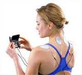
Electrical Stimulation Therapy
Jeffrey Larson, PT, ATC
The history and
origin of electrostimulation, also commonly referred to
as electrotherapy, is unique. The therapeutic benefits
of electricity were not discovered in a laboratory or
clinic and were not a byproduct of someone's
accidentally coming into contact with a bolt of
lightening. In fact, electrotherapy originates as early
as 400 BC from contact with the torpedo fish, which
could produce electric shocks between 100 and 150 volts.
Taken live from lakes and streams and placed on a
painful area of the body, the torpedo fish produced a
series of electric shocks that reduced and controlled
pain.
In the mid 1700s the development of the Leyden Jar,
which is a predecessor to the battery, provided the
capacity to store electricity. As a result, physicians
had more control over where, when, and how much current
could be applied for therapeutic use. The advancement of
the battery in the 1800s further developed
electrotherapy, and in the latter half of the nineteenth
century most physicians in America possessed at least
one electrical stimulator. However, as with any new
medical technology, electrotherapy was not readily
accepted. This skepticism resulted in a decline of
interest in electrotherapy toward the end of the
[nineteenth] century (eMedicine Clinical Knowledge Base,
1996).
In 1965 electrotherapy regained its popularity when
the gate control theory of pain was introduced. This
theory proposed that pain perception depends on the
balance of large- and small-diameter nerve fiber
activity and that an increase in large nerve fiber
activity can potentially "close the gate" to information
going to the brain from small pain fibers. When the gate
is closed, the transmission of pain signals to the brain
is blocked.
Clinical evidence came in 1967 by Wall and Sweet, who
reported that electrical nerve stimulation provided
successful relief of chronic pain. Initially, electrodes
had to be surgically implanted but it was discovered
that current could be sent directly through the skin,
eliminating the need for surgery. This therapeutic
effectiveness in pain relief has led to other
applications of electrotherapy by rehabilitative
clinicians, including treating injured or diseased
muscle and other soft-tissue conditions (Gersh, 1992).
This course reviews cell physiology and the response
of muscle fibers to electrical stimulation, and presents
the principles of electrical stimulation to aid the
healthcare professional in decisions regarding
indications and clinical applications.
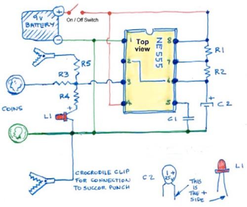
THE PHYSIOLOGY UNDERLYING ELECTROSTIMULATION
Excitable Cell Membranes
The major therapeutic uses of electricity derive from
muscle contractions or sensory stimulation or a
combination of both, so it is important to review the
general physiological effects of electricity on nerve
and muscle tissues. Nerves and muscles are both
excitable tissues, and this
excitability is dependent on permeability of
the cell membrane. The nerve or muscle-cell membrane
regulates the interchange of substances from inside and
outside the cells.
This cell permeability is voltage-sensitive,
producing an unequal distribution of charged ions on
either side of the cell membrane, which in turn creates
a difference in electrical charge between the interior
and exterior of the cell. When this charge occurs, the
membrane is considered to be polarized. The potential
difference between the inside and outside charge is
known as the resting potential because
the cell tries to maintain this difference in electrical
charge as its normal
homeostatic environment.
There is a greater concentration of diffusible
positive ions outside the membrane than within it. The
cell continuously moves positively charged sodium from
inside to outside, and balances this by moving
negatively charged potassium to the inside through a
mechanism called active transport. A
higher concentration of potassium occurs inside the
cell, but the overall charge difference produces an
electrical gradient with positive charge outside and
negative charges inside (Gersh,1992).
Nerve and Muscle Fibers
The nervous system is subdivided into the central
nervous system (CNS) and peripheral nervous system. The
CNS comprises the brain and spinal cord.
The peripheral nervous system consists of nerves
emerging from the brain (cranial nerves) and from the
spinal cord (spinal nerves). Spinal-nerve fibers of the
peripheral nervous system that convey conscious and
unconscious information such as pain are termed
afferent fibers.
Spinal-nerve fibers that control voluntary muscles
are efferent fibers. Both the
peripheral afferent input and central efferent activity
are of interest to the physical therapist employing
electrical stimulation (Noback, 1991).
When an electrical stimulation system is applied to
the soft tissues of an extremity, the system provides
the force to induce ion movement into the tissues and
create an action potential. At the positive electrode
(anode), positive ions are repelled and negative ions
are attracted. At the negative electrode (cathode), the
negative ions are repelled and positive ions are
attracted. As the charged ions move across the
nerve-fiber membranes beneath the anode and cathode,
membrane depolarization occurs.
The cathode is usually the area of the depolarization
and, as a concentration of negatively charged ions
increases, the membrane's voltage potential becomes low
and is brought toward the threshold of depolarization.
The anode makes the nerve cell membrane more positive,
increasing the threshold necessary for depolarization.
In terms of muscle excitation, a twitch muscle
contraction results. This contraction, initiated by
electrical stimulus, is the same as a twitch contraction
coming from voluntary efforts or activity (Prentice,
2001).
THE PHYSICS OF ELECTROSTIMULATION
Alternating and Direct Current
Electrotherapeutic devices used in rehabilitation
generate two different types of current that, when
introduced into biological tissues, are capable of
producing specific physiologic changes. These two
current types are referred to as alternating and direct
current. In alternating current, the
electrons constantly change directions, reversing its
polarity. Electrons flowing in alternating current
always move from the negative to positive pole,
reversing direction when the polarities are reversed.
Conversely, direct current is a
unidirectional flow of electrons toward the positive
pole. However, on most modern direct-current devices,
the polarity and thus the direction of current flow can
be reversed. Electrotherapeutic devices are usually
further classified as being either high-voltage
generators or low-voltage generators. The high-voltage
devices produce waveforms (the visual representation of
the current or voltage) within an amplitude of 115 volts
and are greater and of relatively short duration (less
than 10
msec) (Gersh, 1992).
Pulsed Current
Pulsed current is the unidirectional or bidirectional
flow of charged particles that periodically stop for a
limited period of time before the next event. More
specifically, a
pulse is an isolated electrical event
separated by a finite period of time from the next
event. A constant current source is preferable to a
constant voltage source for most physiologic
applications (Gersh, 1992; Prentice, 2001).
Three types of current.
Galvanic Current
The terms galvanic current and
direct current are often used interchangeably.
Historically, the term galvanic has been used
to describe an uninterrupted direct-current form.
High-volt pulsed galvanic electrical stimulators are
considered to be useful in acute injuries associated
with major tissue trauma accompanied by bleeding or
swelling. Their direct current creates an electrical
field over the treated area that, theoretically, changes
blood flow.
Connected to two pads, galvanic stimulation uses a
positive pad that behaves like ice, causing reduced
circulation to the area under the pad and an associated
reduction in swelling, and a negative pad that behaves
like heat, causing increased circulation and reportedly
speeding healing (Gersh,1992).
Interference Current
Interference current is based on the summation of two
alternating-current signals of slightly different
frequency. This results in current having a recurring
modulation of amplitude, based on the difference in
frequency between the two signals.
TYPES OF ELECTRICAL STIMULATION
Iontophoresis
Iontophoresis, the process of
increasing the penetration of drugs into the skin by
application of an electric current, is commonly used by
physical therapists for the purpose of delivering
anti-inflammatory medications such as corticosteroids.
The groundwork for iontophoresis dates back to the early
1900s, with initial scientific experiments performed by
a researcher named LeDuc.
The majority of units consist of a compact
phoresor that operates with a 9-volt battery
and two wire leads, each connected to an electrode. One
electrode is the drug-delivery electrode intended for
the anti-inflammatory, and the other is used as a
dispersive electrode charged opposite to the
anti-inflammatory ion. When the electrodes contain
solutions of ions, negatively charged anions are
repelled from the cathode into the body and positively
charged cations are repelled into the targeted body area
from the anode.
This effect is specific for ions of the same polarity
as the electrode and, conversely, ions of the opposite
polarity are not transferred into the body. Physical
therapists use iontophoresis based on this penetration
and distribution of ions primarily for controlling and
reducing inflammation. This is applied while minimizing
the systemic concentration caused by circulatory removal
of the desired medication from the targeted area.
Two typical prerequisites for treatment with
iontophoresis are that the medication must be charged
(or modified to carry a charge) and that the
inflammatory process be near the body surface (i.e. a
superficial muscle or tendon rather than a deeper muscle
tendon bursa) (Costello, 1995).
The effectiveness of the ion transport system remains
controversial. For example, some researchers have
proposed that all the material delivered through the
skin with iontophoresis is removed by the subcutaneous
circulation and circulated around the body, providing
little if any local concentration to the intended
region. Conversely, other researchers have shown with
animal studies that ions and other substances do
penetrate and do provide local concentration.
In the physical therapy setting, constant direct
current has been commonly used in iontophoresis
applications. However because of concern over pH
changes, some researchers contend that a method of
producing a more "consistent" constant current should be
used to provide current while the skin resistance is
changing. Because of potential skin charge accumulation
and skin irritation due to pH changes, modulated
currents have been used with success on laboratory
animals. Pulsed currents have proved to be as effective
or more effective in the delivery of small ions. Such
studies indicate the need for physical therapists to
consider and investigate the use of currents other than
the traditional continuous monophasic current for
iontophoresis.
Corticosteroids are the principal drugs used with
iontophoresis in physical therapy because they have an
anti-inflammatory effect and are relatively inexpensive.
Dexamethasone is available in a somewhat more stable
dissolved form and is therefore often used with
iontophoresis. Some clinicians recommend treatments
using a current of 4 mA for 10 minutes. This current is
thought necessary to penetrate into the deeper tissues;
however, treatment times greater than 10 minutes are
less likely to achieve any greater tissue concentration
due to circulatory removal of the medication.
Still other clinicians propose a current of 2.0 mA
for 20 minutes for more superficial areas with a chronic
inflammatory condition. More recent advances in this
technology have introduced a disposable single-use
iontophoresis system with an internal battery and
current limiting circuitry. This method provides a
constant drug delivery for an 80 mA-minute treatment and
can deliver both negatively and positively charged drug
ions. It operates at a low current and is worn for 24
hours to deliver the desired dose. The unit is designed
to begin a treatment as soon as it is hydrated and
applied to the skin, and stop the treatment at
approximately 80 mA-minutes (Morris, 2003; Reena Rai,
2005).
Transcutaneous Electrical Nerve Stimulation (TENS)
Transcutaneous electrical nerve stimulation
(TENS) is one of the most commonly used forms
of electrostimulation for pain relief. Numerous clinical
reports exist regarding the use of TENS for conditions
such as low back pain, myofascial and arthritic pain,
neurogenic pain, and postsurgical pain. The method of
pain reduction produced by TENS is explained by the gate
control theory proposed by Melzack and Wall in 1965. The
"gate" between the level of the spinal cord and the pain
centers of the brain usually is closed, thereby
inhibiting constant nociceptive transmission by way of C
fibers from the periphery to the T cell.
When painful peripheral stimulation does occur, the
information carried by C fibers reaches the T cells and
opens the gate, allowing pain transmission centrally to
the thalamus and cortex, where it is interpreted as
pain. Recall that the gate control theory
postulated a mechanism by which the gate is closed
again, preventing further central transmission of the
nociceptive information to the cortex. The proposed
mechanism for closing the gate is inhibition of the
C-fiber nociception by impulses in activated myelinated
fibers (eMedicine Clinical Knowledge Base, 1996; Gersh,
1992; Noback,1991).
A TENS unit consists of one or more electric signal
generators, a battery, and a set of electrodes. The
units are small and programmable, and the generators can
deliver uninterrupted forms of stimuli with variable
current strengths, pulse rates, and pulse widths. The
preferred waveform is biphasic, which helps avoid the
electrolytic and iontophoretic effects of a
unidirectional current. A variety of newer
transcutaneous or percutaneous electrical stimulation
modalities are emerging as technology advances (Jarzem,
2005).
Interferential Current Therapy (IFC)
Interference current, utilized as
interferential current therapy (IFC), is based
on the summation of two alternating-current signals of
slightly different frequency. This results in current
having a recurring modulation of amplitude, based on the
difference in frequency between the two signals.
When these signals are in phase, they sum to an
amplitude sufficient to stimulate; when the signals are
out of phase, no stimulation occurs. To determine the
stimulation rate of an IFC unit, you must understand
that the
beat frequency of IFC is equal to the
difference in the frequencies of the two signals. For
example, the beat frequency and, hence, the stimulation
rate of a dual-channel IFC unit with signals set at 3400
and 3300 Hz is the difference of 100 Hz.
Interferential current therapy can deliver high
currents compared to other stimulators, and can use 2,
4, or 6 applicators, arranged in either the same plane
for use on regions such as the back or in different
planes in complex regions such as the shoulder
(eMedicine Clinical Knowledge Base, 1996; Gersh,1992).
Neuromuscular Electrical Stimulation (NEMS)
Neuromuscular electrical stimulation (NMES)
is the application of current to elicit a muscle
contraction. The use of NMES in orthopedic and
neuromuscular rehabilitation has grown significantly in
recent years. A nerve action potential may be elicited
either by a command originating in the motor cortex of
the brain or by an electrically induced stimulus at the
periphery. NEMS is addressed below under Stimulating
Muscle Contraction.
Functional Electrical Stimulation (FES)
Functional electrical stimulation (FES)
is another form of electrotherapy that utilizes
electrical currents to activate the nerves that serve
extremities affected by paralysis. This paralysis can be
the result of a spinal cord injury, head injury, stroke
or other neurologic disorders. The goal of FES is to
help restore function in people with disabilities. The
many possibilities for patient management using FES are
beyond the scope of this course. However, a description
of the use of FES for normalizing gait pattern in
included under Stimulating Muscle Contraction, below.
TERMS USED IN ELECTRICAL STIMULATION
Voltage
The electrical force needed to produce a flow of
electrons (current) is called a volt. The volt
is a unit that describes the difference in the
concentration of electrons between two points; the
electrons cannot move unless such a difference exists.
Voltage measures the potential energy
of an electric field that results from the accumulation
of electrons at one point in an electrical circuit and a
corresponding shortage of electrons at another point in
the circuit. If the two points are connected by a
suitable conductor, the potential difference will cause
electrons to move from an area of higher population to
an area of lower population. Commercial current flowing
from wall outlets produces either 115 volts or 220
volts. Electrotherapeutic devices used in injury
rehabilitation are capable of modifying voltages.
Resistance and Impedance
The opposition to electron flow in a conducting
material is referred to as resistance.
Resistance is measured in ohms. The mathematical
relationship among current, voltage, and resistance is
current = voltage/resistance (Gersh, 1992 ; Hayes,
2000).
Impedance is the resistance the
body's tissue to the passage of electrical current. Bone
and fat are high-impedance tissues; nerves and muscles
are low-impedance tissues. If a low-impedance tissue is
located under a large amount of high-impedance tissue,
the current may never become high enough to cause
depolarization.
Waveforms
The term waveform refers to a
graphic representation of the shape, direction,
amplitude, and duration of the current being produced by
the electrotherapeutic device. Both alternating and
direct currents may take on a design of sine, square, or
triangular waveform arrangement, depending on the
capabilities of the electrostimulation device producing
the current. The basic difference between alternating
and direct current is that for each shape the
alternating current reverses direction one time in each
cycle; the direct current does not reverse direction. If
the modality has capabilities of automatically reversing
polarity, a direct current will elicit the same
physiological response as an alternating current (Hayes,
2000).
Essential Parameters
Electrostimulation parameters for therapeutic
applications are defined in terms of duration, strength,
frequency, on-off time, rise-fall time, and polarity.
Some parameters are time-dependent; pulsed current is
described by special time-dependent properties of the
pulse. The term phase
describes the current moving in one direction for a
predetermined period of time. The pulse waveform may be
monophasic or biphasic. Monophasic refers to the current
located on one side of the baseline, whereas biphasic
current is present on both sides of the baseline.
Phase duration is the time elapsed
from the beginning to the end of one phase.
Pulse duration, also known as "pulse width," is
the time elapsed from the beginning to the end of all
phases in one pulse (Figure 2). The rise time
measures the time for the leading edge of the phase to
increase from the baseline to the peak amplitude of
phase (Figure 2). The fall time is the time for the
terminal edge of the phase to return to the zero
baseline from the peak amplitude of the phase.
Frequency is the repetition rate of the
waveform expressed in pulses per second or cycles per
second. Both alternating and pulse currents are
described by frequency-dependent properties.
Graphs showing pulse
duration and rise time.
For clinical purposes, pulse and alternating currents
can be varied within a specific time frame. Pulse
duration, phase duration, and frequency may also be
modulated. Ramp, or surge, modulations are increases or
decreases in the phase charges over time. A
train is a continuous repetitive sequence of
pulses or cycles of pulsed current. A burst
is an interruption in a train separated by an
inter-burst interval. The duty cycle is the ratio of
one-time to total-time of trains. The duty cycles are
generally expressed as a percentage. For example, if the
pulse duration of the waveform is 25 msec and the period
is 100 msec, the duty cycles are 25/100 or 25%.
Also of clinical importance is the intensity or
strength of the electrical stimulus. Increasing the
intensity or strength of the stimulus allows the current
to reach more deeply into the tissue. The depolarization
of more fibers is then accomplished by depolarizing
higher threshold fibers within the range of the first
stimulus. Additional fibers with the same threshold but
located deeper to the structure are depolarized by
deeper spread of the current (Gersh, 1992; Hayes, 2000).
USING ELECTROSTIMULATION DEVICES
The Electrodes
Electrodes are fabricated of electrically conductive
material that is used to transfer electric charge to
biological tissue. Similar to the electrodes of
recording systems such as electromyography (EMG),
therapeutic stimulating electrodes may be used on the
skin surface or percutaneously; however, percutaneous
electrodes are less commonly used in therapeutic
electrostimulation for rehabilitation purposes.
The percutaneous type of electrode system comes into
direct contact with body fluids and has lower impedance
than surface electrodes. Surface electrodes normally
require some type of skin preparation to reduce skin
impedance, as well as the use of an electrolyte couple
medium. An electroconductive gel applied between the
electrode and skin serves to reduce skin impedance. More
recent electrode designs are pre-packaged with both a
conductive gel and an antibacterial component. The
couple medium needs to be kept moist because skin
irritation can occur in as many as 33% of patients, due
partly to drying out of the electrode gel.
Electrode shapes can be round, oval, or rectangular
and can also be custom fitted; sizes vary from 3 to 5
inches in diameter. With consistent electrode
conductivity, current density is inversely proportional
to the electrode size. While the contact area of the
stimulating electrode decreases for specific current
intensity, the current density increases. This
phenomenon explains why the smaller electrode in a
monopolar configuration is selected to be the active
electrode, while the larger electrode serves as the
dispersive or reference electrode. Because electrode
size is directly proportional to the current level,
larger electrodes have lower impedance—that is, for a
given voltage a larger electrode provides a greater
current level (Gersh, 1992; Hayes, 2000).
Placement Techniques
Guidelines for placing electrodes are the same no
matter what protocol is used for stimulation of sensory
nerves. These guiding principles are designed to help
clinicians select the appropriate sites for electrode
placement. The TENS applications use electrodes sized
similarly to other electrostimulating devices; they are
placed according to a pattern and moved about in a
trial-and-error manner until pain is decreased.
Electrodes may also be placed directly on or around
the painful area, and over specific
dermatomes, myotomes, and sclerotomes
corresponding to the painful region. Electrodes may also
be placed near a spinal cord segment that innervates the
area that is painful. Another option to the clinician is
the peripheral nerves that innervate the painful area.
The peripheral nerves can be stimulated by placing
electrodes over sites in which the nerve becomes more
superficial and is stimulated more easily.
The orientation of electrodes used in therapeutic
electrical stimulating systems can be monopolar,
bipolar, or quadripolar. When applying a
monopolar orientation, the electrode of the
stimulating circuit is placed over the target tissue and
is referred to as the active electrode. This location is
where the greatest effect is desired; a second, larger
dispersive electrode is placed some distance from the
active electrode.
In the bipolar configuration, a
dispersive electrode is not required because two
electrodes from one circuit are placed over the target
tissue; one electrode is positive and the other is
negative. In the quadripolar design,
electrodes from two or more circuits are positioned so
that currents intersect; this type of electrode
placement may be used for interferential stimulation
technique (Gersh, 1992; Hayes, 2000).
APPLICATIONS
Managing Pain
Electrical stimulating systems are commonly used in
the treatment of acute and chronic pain. The most
successful application of
TENS continues to be the control of
postoperative incision pain. It is most effective when
the postoperative pain is confined to a small area, is
well-defined, and is generally self-limiting as to time,
course, and severity. Clinicians often expose patients
to TENS during instruction about stimulus parameters to
be used postoperatively. During this pre-operative
instruction, attention should be given to the patient's
own sensory requirements (personal feedback) regarding
selection of stimulation parameters.
The concurrent use of analgesic medication to address
postoperative pain may skew the need for TENS. In
addition, medication history may be a compounding factor
in evaluating the effect of TENS on postoperative pain.
Researchers evaluating the effect of TENS on patients
following a laminectomy noted that TENS was most
effective for managing pain in the drug-inexperienced
patients—described in the study as patients who had had
no more than 2 weeks' narcotic medication in the 6
months prior to surgery. Patient cost benefits from the
use of TENS include reduced narcotic intake and a
potential decrease in incidence of depression. Early
mobility, fewer instances of postoperative pulmonary
complications, and, in some cases, a reduced length of
stay in the ICU are also possible benefits of TENS.
Other electrical stimulating alternatives to
conventional TENS for pain control include the
application of interference current. Recall that
IFC consists of waves of constant amplitude
and slightly different frequencies from two independent
low-voltage AC currents. The waves are superimposed,
resulting in higher-amplitude wave produced secondary to
the summation of current values at that specific point
in time.
Some clinicians who advocate interference current for
treatment of muscle pain contend that IFC penetrates
deeper than other forms of TENS based on the rationale
that skin impedance decreases in response to higher
frequencies of alternating current.
Interferential current therapy may be applied in such
manner that combined "beat" characteristics produce a
stimulus that corresponds to those produced by brief,
intense TENS. Setting the beat frequency between 100 and
120 bps produces beat durations of between 10 and 8
msec, respectively. By setting amplitude to the maximum
tolerable output, a strong continuous tingling
paresthesia combined with mild to moderate muscle
facilitations is produced. Using this method of exciting
motor and sensory nerve fibers at the same time, pain
reduction can be induced within 15 minutes (ProMax,
2007).
Determining electrode sites for optimal stimulation
when treating acute and chronic pain is not specifically
documented. Electrode placement varies but it is
commonly at the site of pain, adjacent to the spinal
column at the spinal nerve root, or over the course of
the peripheral nerve serving the painful region. The
selection of preferred stimulation sites is made once
the nature, location, and structural source of pain have
been determined. The spinal-cord segments and peripheral
nerves innervating that structure are identified on
initial examination.
Preferred stimulation sites along the innervating
structures may be located using motor points, trigger
points, or acupuncture-point charts or tables, or by
palpation and knowledge of the anatomy. Clinicians can
also search for regions of high conductivity by holding
one electrode of a TENS circuit and giving the patient
the second electrode. After turning on the electrical
current, clinicians places their index finger on the
patient's skin overlying the course of the peripheral
nerve that innervates the painful region. As the finger
is moved along the skin, the patient reports sites at
which the electrical stimulation is perceived most
acutely. These are points of low skin resistance and
they are thus more susceptible to electrical stimulation
(Gersh, 1992; Hayes, 2000; ProMax, 2007).
Reducing Edema
Electrical stimulation can be used for various types
of edema reduction. Traumatic edema resulting from the
disruption of blood vessels often accompanies
musculoskeletal injuries such as acute strains and
sprains. Voluntary muscle pump activity or muscle pump
facilitation through electrical stimulation may be
effective in lymphatic and venous drainage and thus aid
in resolution of posttraumatic edema.
The second approach for reducing traumatic edema
through electrostimulation employs sensory-level
stimulation that does not result in muscle contraction.
Clinicians theorize that perhaps an effective current in
managing edema would be a low-intensity, continuous,
unidirectional current that would be expected to have
the appropriate polar effects dependent upon the
specific polarity used for treatment (Gersh, 1992;
Hayes, 2000).
Stimulating Muscle Contraction
USING NMES
Neuromuscular electrical stimulation (NMES) is the
application of current to elicit a muscle contraction.
As mentioned earlier, the use of NMES in orthopedic and
neuromuscular rehabilitation has grown significantly in
recent years. A nerve action potential may be elicited
either by a command originating in the motor cortex of
the brain or by an electrically induced stimulus at the
periphery.
The optimal size of the electrode is based on the
desired muscular response, the size of the target muscle
or muscles, and the chosen electrode placement. Larger
electrodes are effective in generating torque from large
muscles or groups of muscles that contract together; for
example, a 5 cm x 10 cm electrode may be used to
stimulate the quadriceps and hamstring muscles.
When small individual muscle stimulation is desired,
a small electrode that increases the current density is
placed over the motor point of that muscle; a second,
larger reference electrode is often placed distally over
the tendinous portion of the muscle. The second
electrode provides a return path for the current but has
inadequate current to cause significant polarization of
excitable tissue under the electrode.
Recall that monopolar stimulation occurs when one
electrode is placed over a motor point with the other
placed at a site away from that area, while bipolar
stimulation describes placement of both electrodes on
the muscle or muscle group to be activated. Bipolar
placement tends to be used most often in neuromuscular
electrostimulation, because, for a given intensity of
stimulation, more current reaches the muscle to be
stimulated.
For optimal bipolar positioning, the clinician should
place the muscle at resting length or in a slightly
lengthened range, avoiding any close-packed positions of
the limb or joint. Isometric contractions are used to
achieve greater tension and should be applied at several
points in the range of motion. Greater tension is built
with higher frequencies but it can cause fatigue, and a
frequency of 50 pulses per second (pps) provides as much
as the higher frequencies. The amplitude is increased
until strong maximal contraction is obtained; however,
patient tolerance is the guide and stimulation should
not be painful.
A rest cycle that is 5 to 6 times as long as the hold
cycle allows the muscle adequate time to recover between
contractions and produces same amount tension on each
subsequent contraction. A hold time of 6 to 10 seconds
builds optimal tension and 8 to 10 contractions in a
single treatment session are sufficient for
strengthening. Strength gains are likely to begin
peaking about 20 to 25 sessions. Treatment should be
provided daily, or at least every other day (Gersh,
1992; Hayes, 2000).
Electricity has been used to stimulate denervated
muscle for over a century, yet the success rate is
unclear; while this form of treatment has been shown to
be effective in laboratory animals, studies are not
consistent regarding its effectiveness in humans. The
rationale for electrically stimulating denervated muscle
is to exercise the muscle in an attempt to maintain it
in a healthy condition while the injured axons
regenerate and re-innervate the muscle.
Following the denervation of a muscle, the tissue
undergoes a number of changes that are physiologic,
biochemical, and anatomic. Progressive muscle fiber
atrophy is the most obvious change, and the
neuromuscular junctions also begin to degenerate. To
achieve successful treatment of denervated muscle,
electrical stimulation should be initiated as soon after
the injuries as possible. In some cases, atrophy that
has already occurred cannot be reversed. A stretched
muscle produces more tension, and greater tension is
produced with isometric contractions when they are
feasible.
Denervated muscle requires a stimulus of long
duration, and greater than 100 msec is recommended.
Alternating current of 5 Hz has been known to provide a
phase duration of 100 msec and to be preferred for the
patient's skin. The use of continuous direct current
that is manually interrupted can also provide a desired
response. If direct current is used, the cathode should
be the stimulating electrode. A 100 msec phase duration
can be delivered only 10 times per second, assuming no
rest between stimuli.
The amplitude is increased until a strong contraction
is obtained, within the patient's tolerance, and
sustained for at least 2 seconds. A period of rest
follows the contraction that is 5 times as long as the
length of the contraction. Permit 10 to 25 contractions
per session which is to be completed 3 times a day with
at least 10 minutes of rest between sessions. Treatment
can be uncomfortable for the patient and consistent
encouragement may be needed to ensure continued
compliance at these parameters (Gersh, 1992; Hayes,
2000).
USING FES
Functional electrical stimulation (FES) is another
form of electrotherapy that utilizes electrical currents
to activate nerves that serve extremities affected by
paralysis. The goal of FES is to restore function in
people with disabilities. There are gait deviations
associated with orthotic management, and
electrostimulation that is timed to coincide with the
individual gait pattern can be a useful addition to many
gait training programs. This can take form of a
conventional ankle orthosis made of plastic/metal and
leather with the addition of electrical stimulation
using a remote control switch.
When the clinician chooses electrostimulation as an
orthotic substitute, the timing of the stimulus during
ambulation is usually controlled by a pressure-activated
heel switch. As a result, corrective positioning
provided during the swing-through phase will be
terminated during stance. The corrected positioning for
stance will be terminated during swing-through,
producing a more normal gait pattern.
If dorsiflexion during swing-through is the goal of
stimulation, there should be no interference with
push-off during stance, because weight-bearing on the
heel switch during stance would inactivate stimulation
of the dorsiflexors. Similarly, where knee extension is
stimulated for stance stability, there would be no
interference with knee flexion swing-through because
non–weight-bearing on the heel switch at end of stance
would inactivate stimulation of the quadriceps muscle.
When electrostimulation is used to provide
dorsiflexion during swing, the electrical stimulus stops
upon weight-bearing, thereby allowing the ankle to move
freely into plantar flexion from heel-strike to
foot-flat. As a result, no increase in flexion is
produced at the knee. This can be important for the
hemiparetic patient, where instability is often a
problem.
Relatively small changes in electrode position can
noticeably influence the effect in joint positioning.
When electrostimulation as an orthosis is used outside
the clinic, it is essential to have the patient or
attendant reliably duplicate effective placement and be
aware of changes in joint positioning during the wearing
period.
Because the electrostimulation protocol is more
complex than a functional orthosis, there is a greater
chance of mechanical problems and/or human error. Safety
is a primary concern and any human error can result in a
change in joint positioning, placing the patient at risk
of falling. These factors are much less problematic
during supervised use of electrostimulation in the
clinical setting (Gersh, 1992 ; Hayes, 2000).
Improving Circulation
Circulation can be improved with
NMES by increasing metabolic demand through
sporadically activating the muscle pump around the
circulatory network. In this application, a low
frequency of 20 to 30 pps has been shown to be most
effective. A contraction of only 10% to 30% of maximal
effort is sufficient. Duty cycle should be one that is
not fatiguing; treatment may be given for 10 to 30
minutes and as frequently as is comfortable for the
patient (Prentice, 2001; Gersh, 1992; Hayes, 2000).
Healing Wounds
There is now an ample body of evidence supporting the
use of electric stimulation for wound healing.
Guidelines for the use of stimulation have not been
determined for all conditions, yet enough information
exists to guide the clinician to a reasonable plan of
care. Although not conclusive, a body of evidence
suggests that polarity is an important treatment issue
for both acute and chronic wounds.
RATIONALE
Literature on wound healing describes the body as
having its own bioelectric system, which influences
wound healing by attracting the cells of repair and
changing cell membrane permeability. When there is a
rupture in the skin, a current is generated between the
skin and inner tissues that continues until the skin
defect is repaired. Healing of the injured tissue is
impeded or will be incomplete if these currents no
longer flow while the wound is open. A rationale for
applying electrical stimulation is that it mimics the
natural current of injury and will initiate and/or
accelerate the wound healing process.
Also of clinical importance is the theory that a
moist wound environment is required for the bioelectric
system to function. By keeping the wound moist with
normal saline, the ideal electrical charge is maintained
and dressings such as hydrogels and occlusive dressings
can help promote the body's own bioelectric system by
keeping the wound moist (Gogia, 1995).
Clinicians understand that wound debridement is
enhanced if the tissue is solubilized with enzymatic
debriding agents. Electric stimulation using negative
current has been shown to solubilize clotted blood,
whereas the positive electrode has been found to induce
clumping of leukocytes and formation of thrombosis in
the small vessels. This helps explain why clinical
observations demonstrate that hemorrhaging at the wound
margin is dissolved and reabsorbed following application
of high-volt pulsed current (HVPC) with
the negative pole.
Treatment parameters differ considerably in recent
studies of successful healing. Typically the active
electrode, which is one-fourth the size of the dispersal
electrode, is positioned directly over the wound. To use
this form of stimulation, electrodes may need to be cut
to the appropriate sizes.
Placement of the dispersal electrode in relation to
the active electrode is another area of controversy.
Some researchers recommend the placement of dispersive
electrodes closely proximal to the origin of the spinal
nerve; in other words, while treating wounds with
negative polarity, the dispersive or positive electrode
should be placed close to the spinal cord relative to
the negative electrode.
Electrical stimulation affects each phase of wound
healing differently, beginning with the inflammatory
phase that initiates the wound repair process. In this
phase, increasing blood flow can help in the removal of
debris by way of phagocytosis; in addition, by
increasing blood flow, electrical stimulation enhances
tissue oxygenation.
Next, electrical stimulation has been determined to
promote the proliferation phase by stimulating the
fibroblasts and epithelial cells needed for tissue
repair. Membrane transport is improved, which supports
the body's natural current.
Eventually, in the proliferation stage, better
collagen is produced, which helps in the stimulation of
wound contraction. Clinical studies suggest settings for
the proliferation phase with the polarity as negative,
pulse rate at 100 to 128 pps, intensity of 100 to 150
volts, and a duration up to 60 minutes, once daily, 5 to
7 times per week.
In the later phase of epithelialization,
electrotherapy can stimulate epidermal cell reproduction
and migration, helping to produce smoother and thinner
scar tissue. Settings during the epithelialization phase
include alternating the polarity every 3 days—for
example, 3 days negative followed by 3 days positive.
Pulse rate is recommended at 64 pps, an intensity of 100
to 150 volts, and a duration of 60 minutes, 5 to 7 times
per week.
Based on scientific rationale from early studies, the
application of electrical stimulation using direct
current reported long treatment times of 20 to 40 hours
per week. Four controlled clinical studies and three
uncontrolled studies with
HVPC reported a mean healing time of 9.5
weeks with 45- to 60-minute treatments, 5 to 7 times per
week (Sussman, 2000).
PREPARING THE PATIENT FOR WOUND TREATMENT
Having the supplies ready before removing the wound
dressing saves time and helps avoid unnec