Outer Ear,middleear,inner ear,illustrated diagram
Scala vestibuli ,Scala tympani, Reissner's membrane,Round window
continued from the Brain Page of Nervous System
Contents
neurons and Nervesneurotransmitter
The Brain & Spinal Cord
Cranial Nerves
Peripheral Nervous System
Autonomic Nervous System
Senses: Eye diagrams, Hearing, Smell,Taste, Taste & Tongue Sensation,Balance
Memory , Memory types, Creation of Memory,
Higher Functions
Altered States
- Hearing (see location of the various components in Figure 09):
- Outer Ear -
- Pinna - This is the external flap for collecting sound wave.
- Auditory canal - The opening of the auditory canal is lined with fine hairs and sweat glands. The modified sweat glands secrete earwax to guard the ear against the entrance of foreign materials, such as air pollutants.
- Middle Ear -
- Tympanic membrane (ear drum) - It pushes a set of three small bone (the ossicles) against an inner membrane.
- Ossicles - The three parts in this structure are: hammer, anvil, and stirrup. These components multiply the sight vibration of the sound by about 20 times. When the stirrup strikes the oval window, the pressure is passed to the fluid within the inner ear.
- Eustachian tube - It extends from each middle ear to the nasopharynx and permit equalization of air pressure. Chewing gum, yawning, and swallowing in elevators and airplanes help to move air through the eustachian tubes upon ascent and descent.
- Inner Ear - Whereas the outer ear and the middle contain air, the inner ear is filled with fluid. Anatomically speaking, it has three areas: the first two, the semicircular canals and a vestibule, are concerned with balance; only the third, the cochlea, is concerned with hearing. It is a very delicate and sophisticated organ about 1 cm3 in size.
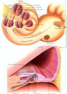
- Scala vestibuli - The incoming vibration (red arrows in Figure 14) spirals along this structure toward the apex of the cochlea. At the apex, a small gap allows the wave to pass into the scala tympani.
- Scala tympani - The wave travel down (blue arrows) in this spiraling tube toward the round window.
- Round window - It acts as a pressure relief membrane dissipating the vibrational energy.
- Organ of Corti - This structure is set onto the basilar membrane which forms one of the arms of the Y inside the cochlea (see Figure 14). Pressure changes in the fluid outside the scala media create vibrations in the basilar membrane and in the Reissner's membrane, which forms the other arm of the Y. The vibrations in the scala media and the tectorial membrane shake the hair cells, which convert the vibrational energy into electrical nerve impulses. The signals are channeled by nerve fibers along the organ of Corti to the main cochlear nerve, and then to the temporal lob of the brain for processing into the sounds that we perceive.


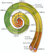
According to the frequency of the sound wave, different parts of the basilar membrane along the organ of Corti are set into motion. In general, low-pitch sounds make the apex of the cochlea vibrate while high-pitched ones cause most vibrations near the base of the cochlea. Figure 15 shows such frequency distribution along the length of the cochlea for both the incoming and outgoing waves. The strength of nerve signals also depends on the volume of the sound. This is interpreted by the brain as loudness. It is believed that tone is an interpretation of the brain based on the distribution of hair cells stimulated.Figure 14 Cochlea
[view large image]Figure 15 Sound Wave
[view large image]
Continued to Taste and Smell page
Scala vestibuli ,Scala tympani, Reissner's membrane,Round window