God is our Guide                 Number 1 site for helping reverse diseases on Planet Earth
cidpUSA Foundation

 cidpusa.org   


      Home
      Diagnosis
      Treatment
      Pathology
      Variants
      CIDP info
      Fibromyalgia
      IVIG
      Diet anti-inflammatory
      Burning  Feet Home
      Services Page
      Chronic Fatigue
      Autoimmune diseases
      Prognosis
      Bible healing
      Celiac disease
Bible page

Autoimmune self attack

What is autoimmune

Autoimmune types

Autoimmune Guide

 Autoimmune-Epidemic

 Autoimmune & women

Autoimmunity secrets

Autoimmune inflammation

Auto-diseases plan

Autoimmune Risk

Autoimmune Rx

Autoimmune anemia

Autoimmune Ear

Autoimmune Thyroid

Autoimmune Fiber

Autoimmune Muscle

Autoimmunity summary

Autonomic Small Fiber

Myasthenia Holistic

Polymyositis

Dematomyositis

Poly Dermato

Myositis

Myasthenia alternative

Myofacial pain

Myopathy

Fibromyalgia

Inner Ear Disease AIED

 
  Natural Makeup
  Neck Pain
  Ocular Female diseases
  Chronic fatigue syndrome
  Osteoporosis
  Women Heart Attacks
  Breast Size & Disease
  Female Sex Disease
  PARKINSON
  Memory problems
  Breast Lymph Drainage
  Kidney stone Buster
 Bras cause breast cancer
  Skin repair Clinic
 Pandas
  Hepatitis


Risk of heart disease & stroke 

Depression and breast cancer

Kidney stone removal 

Alopecia general

Personality

Eye Clinic

Skin hair nail spa

Memory clinic

Depression & anxiety

Addiction  & Drug Rehab

Sexual  disorders Clinic

Parkinson Clinic

Epilepsy Clinic

Pain Clinic

Bone disorders clinic

Joint disorder clinic

Skin repair clinic

Gene Manipulation

Neurology Clinic

TMJ Clinic

Sex in autoimmune disease

Reduce  your weight

Antibiotics

 Want more sex

 Vitiligo 1

 Vitiligo 2

Eliminate risk of heart disease & stroke 

Memory clinic

Sex in autoimmune disease

Reduce weight

Drug reaction prevention

Prevent Osteoporosis

Some rheumatic disorders

Alopecia treatment

Bald Facts

Alopecia

Areata Alopecia

Hair chemicals

Hair Growth

Hair of dog technique

Hair rejuvenation

Nail Fungus

SESAME SEED OIL

Facial  cleaner

oil pulling

 Reading disorders

 Best New Diet

 DHEA Fountain of Youth

DHEA levels and cognitive functions

Parkinson

Diabetise-2

Do not give tots cold medicine 

neurological effects of CIDP

Body goes against the grain

Celiac disease Info

More on Celiac disease

Anemia and celiac disease

Home for autoimmune disorders, information about autoimmune diseases

 Sex clinic

Lahore Clinic page

Everything about IVIg, Home to IVIg

Home to autoimmune diseases, causes, treatment, cure, e-book

Fatty acids in autoimmune diseases

News

Multifocal neuropathy

Dermrmatomyositis

 
 Link to Articles

 www.cidpusa.org/P/ivig.htm


 Information on Taste

  For a simple guide to treatment of autoimmune disease by alternatives please read our e-book
  •  

   See our services section for help and contact information.

continued from the  Brain Pageof Nervous System

Contents

Neurons and Nerves
neurotransmitter
The Brain & Spinal Cord
Cranial Nerves
Peripheral Nervous System
Autonomic Nervous System
Senses: Eye diagrams, Hearing, Smell, Taste, Taste & Tongue Sensation, Balance
Memory , Memory types, Creation of Memory,
Higher Functions
Altered States

[Top]

Continued from Brain


 Continue to next page the cranial nerves
 

  • Continue to taste

    Taste (see location of the various components in Figure 09):

     
  • Tongue - Embedded within the surface of the tongue are four types of taste receptors localized in specific regions on the
    Tongue Papillae 
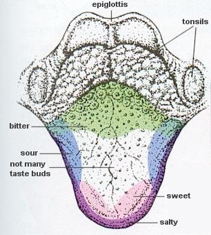
tongue (see Figure 19). Each detects a different class of chemical: sweet (sugars), sour (acids), bitter (complex organics), and salty (salts). The "hot" sensation of foods such as chili peppers is detected by pain receptors, not chemical receptors. But a report in 2006 reveals that contrary to popular belief, there is no tongue map. Responsiveness to the five basic modalities - bitter, sour, sweet salty and umami (a Japanese word

    Figure 19 Tongue
    [view large image]

    Figure 20 Papillae [view large image]


    meaning the savory or meaty taste of amino acids) is present in all areas of the tongue.

     
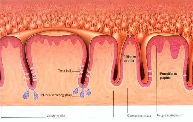
  • Papillae - The papillae are those small elevations visible to the naked eyes. There are three types of papillae located from the back of the tongue toward the tip. Filiform papillae are generally conical or pointed; fungiform papillae are flat-toped; vallate papillae are larger with an outer groove (see Figure 20). Many taste buds lie along the walls of the papillae. Isolated ones also are present on the palate, the pharynx, and the epiglottis.
    Sense of Taste
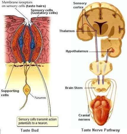
  • Taste buds - The tasting, or gustatory, cells in the buds have hairy tips which detect chemicals in solution (secreted by the gland at the bottom of papilla). When stimulated by flavor molecules, these cells generate nerve signals, which they send to the taste center on the brain's cortex, and also to the hypothalamus, which is concerned with appetite and the salivating reflex.
  • Taste nerve pathway - The nerve signals are carried by three nerves in each side of the tongue (cranial nerves) to a small part of the medulla (brain stem). The signals then travel to parts of the brain, such as the hypothalamus, the thalamus, and the gustatory part of the sensory cortex - the "taste center", where the signals are interpreted (Figure 21). The thalamus acts like a relay station, shunting the data onto appropriate cortical areas for processing. The sense of taste tells us what is good to eat. It evolved to pick out sweet, ripe fruits and energy-packed sugars
  • Figure 21 Sense of Taste
    [view large image]

    and starches. Likewise, taste is is extremely sensitive to bitter flavors, because many poisonous berries, fruits and fungi are bitter-tasting.

     
      Sensations (see location of the various components in Figure 09):

       
    • Skin - Skin has a thin epidermis, which is mainly for protection, and a thicker dermis below. In addition to small blood vessels and sweat glands, it has tiny nerve endings in the various type of touch receptors (see Figure 09).
    • Receptors -
      • Bulb of Krause - These are multi-layered capsules with many branched nerve endings. They are quick-change mechanoreceptors, triggered by rapid alterations in shap caused by pressure or vibrations, and may also help us to feel extreme cold.
      • Free nerve endings - They have a treelike branching system of naked nerve fibers. They are the most common sensory endings in the skin and detect just about anything - light touch, heavy pressure, heat, cold, and importantly, pain. Slight stimulation of these nerve endings may elicit the sensation that is known as itching.
      • Meissner's endings - They are found in the uppermost part of the dermis, especially on the hands, feet, lips, and inner surfaces of the eyelids. They are shaped like eggs and are both quick- and slow-change mechanoreceptors, detecting light touch and vibrations.
      • Merkel's endings - They are like tiny disks stuck in the underside of the epidermis, where they feel slight changes in its shape, thereby detecting light touch. They are both quick- and slow-change mechanoreceptors.
      • Pacinian endings - They have layers like an onion and are sited deep in the dermis. They pick up heavy pressure and also fast vibrations, such as those from a tuning fork.
      • Ruffini endings - They respond to sustained stress or gradually altering shape. This means that they are slow-change mechanoreceptors. They are found mainly in hairy skin and are sausage- or spindle-shaped. It is thought that they may also detect extreme heat.
    • Proprioceptors - The sense of position and movement of limbs is dependent upon receptors termed proprioceptors (Figure 22a). They are located in the joints and associated ligaments and tendons that respond to stretching, pressure, and pain. Nerve endings from these receptors are integrated with those received from other types of receptors so that we know the position of body parts.
    • Sensory nerves - Nerve impulses may reach the somatosensory cortex for analysis before a response is decided. These result in voluntary actions - a deliberate response. Sometimes the stimulus require immediate action (such as from the burning sensation), a reflex action is taken without the conscious control of the brain. These are the involuntary actions directed by the spinal cord. We only become aware of them when other impulses are sent to the brain to "inform" what has happened. The path which impulses travel along during a reflex action is called a reflex arc. Not all the body parts
      Proprioceptors Homunculus receive the same attention of the brain. The relative importance is often represented by mapping over the length of the sensory or motor cortex. These cortical maps (Figure 22b) are not drawn to scale; instead they are variously distorted to reflect the amount the neural processing power devoted to different regions. This accounts for the grotesque appearance of the human body in the homun-culus, which is a translation of the body's sensory map into the human form.

      Figure 22a Propriocep-tors [view large image]

      Figure 22b Homunculus
      [view large image]

       
    Balance

     
      Balance is an ongoing process that keeps our two-legged posture stable. Four main sets of sensory input are involved:

       
    1. Information from the skin is important, especially from the touch and pressure sensors on different parts of the feet, which tell the brain if you are leaning. This sense is not available in a free falling environment such as in a spacecraft.
    2. Eyesight is used to judge verticals and horizontals to which your body should be parallel and at right angle respectively.
    3. The body's proprioceptive sense of stretch in muscles, tendons, and joints tell the brain about the positions and angles of the arms, legs, torso, and neck.
    4. The sensory parts dedicated to balance is located deep inside each inner ear, next to the cochlea (see Figure 09). These parts are known collectively as the vestibular apparatus and are part of the same network of fluid-filled chambers as the cochlea. They consist of the utricle, the saccule, and the semicircular canals (Figure 23a). In certain parts of their linings are tinny hairs, whose roots are embedded in lumpy crystals or gels. The crystals or gels are attracted downward by gravity, and they are also pushed to and fro by the fluid in the chambers, which swirls as the head changes its position.
    Balance
      The functions of the these organs are shown in Figure 23a:
       
    • (a) The ampullae of the semicircular canals contain hair cells with cilia embedded in a gelationous material.
    • (b) When the head rotates, the material is displaced and the bending of the cilia initiates nerve impulses in sensory nerve fibers for maintaining dynamic equilibrium.
    • (c) The utricle and saccule are sacs that contain hair cell with cilia embedded in the gelationous material.
    • (d) When the head bends, otoliths are displaced, causing the gelationous material to sag and the cilia to bend. This initiates nerve impulses in sensory nerve fibers for maintaining static equilibrium.

    Figure 23a Balance
    [view large image]


    The vestibular nerve feeds its information chiefly to the cerebellum and to four structures in the medulla known as vestibular bodies. Using these data, as well as input from the other three sensory sources, the brain works out what to do, usually subconsciously.

     
    Neuromast It turns out that such structure of hair within gel to detect disturbance has been around hundred of million years in the shark and fish (Figure 23b). This is the neuromasts embedded in the skin of fish. They give the fish information about the flow of water. Amphibians and reptiles have a simple uncoiled inner ear. Jawless fish has only one semicircular canal instead of three in mammals (for detecting three dimensional movement). Ultimately, it is the Pax 2 gene that give rise to these structures. It is also known that the Pax 6 gene is responsible for the development of eye. The connection to ancient creatures goes even deeper when it is

    Figure 23b Neuromast
    [view large image]


    found that the box jellyfish carries a gene which is the combination of Pax 2 and Pax 6.
    The box jellyfish is an amazing animal with more than 20 eye pits and many eyes very similar to ours. They seem to double for ears as well.

    [Top]


    Memory

    Types of Memory As shown in Figure 03a, the ability to modify our behaviour in response to life's experiences is shared by all animals including the bacteria E. coli. Such feat requires the brain's willingness to learn. Learning results in the formation of memories and in humans this process reaches its most sophisticated form, allowing us creatively to associate different reflections on the past, to generate new ideas, and most importantly to acquire language as a medium of expression and communication. Memory requires the brain to be physically altered by experience and it is this remarkable property that makes thought, consciousness, and language possible. The basic mechanism of memory formation is highly conservative over

    Figure 24a Memory Classification
    [view large image]

    billion years of biological evolution. The difference in humans is that we have a lot more of the stuffs. There are about 100 trillion synaptic connections in our brain.

     
    Memory There are many ways to classify the memory. The concept of explicit and implicit memory refers to whether or not the recollection is produced consciously and intentionally. While the scheme of declarative and nondeclarative memory depend on the retrieval that can be declared verbally or not. Associative memory is triggered by clues; nonassociative memory can be habitual or sensitive. There are also short term and long term memory. One of the classification schemes is shown in Figure 24a. Table 06 is an attempt to put them all together. In the table, the declarative, and the procedural memory are explicit with the rest of nondeclarative memories being implicit. Only the working memory belongs to the category of short term memory fading away in hours, while the others are long term, and available for retrieval in years. Figure 24b shows the components,

    Figure 24b Types of Memory
    [view large image]

    locations, and pathways for many types of memory.


    continue to TMemory types

  •   

       World Wide Consultation by Internet


    Tremor

    Top Foods

    Treatment of Myasthenia

    Trends in medicine 

    Ulcerative colitis 

    Ultrasound

    Everything about IVIg, Home to IVIg

    Fatty acids in autoimmune diseases

    Small fiber neuropathy 

    Depression and breast cancer

    Dermatomyositis

    Fighting with spouse gets good results!

    Vitamin D  deficiency causes high BP

     Mobile phone Use Increases Tumour Risk

    New Psoriasis Rx       LasiK Dangers

    No cold meds to kids, says FDA

    Calcium supplements cause stroke in women

    Vitamin D extends life   Breast Size & disease

    More Young Women Dying Of Heart Disease

    By pass heart surgery

     Reverse Chronic Fatigue syndrome & Fibromyalgia, 


    BPA in infant bottles a concern

    Want more sex

     Boy or Girl   Autism & Mercury

     Before getting child vaccination read this

     Parents rejecting dangerous vaccine 

    Botox causes deaths also in children

    Autoimmune reaction kills teenager in breast surgery

    Parkinson Prevention    Aids Vaccine alert

    Folate Deficiency Triples Risk of Dementia

    DHEA levels and cognitive functions

    SESAME SEED OIL

    Facial  cleaner

     Quran page