|JULY 4, 2020
Electromyographic (EMG) examination Myopathy, Anterior horn cell disease, Neuropathies, Neuromuscular transmission disease.
3) Electromyographic (EMG) examination
This test consists of two parts: Nerve conduction studies and ElectroMyoGraphy
N.C.V. or Nerve conduction study examines nerve disease. While E.M.G. or Electro Myography examines Muscle disease.
a) Nerve conduction studies
Since there are few pure motor nerves to study, motor nerve conduction recording electrodes are placed over a distal muscle (i.e. thenar muscle group). The appropriate nerve is then stimulated electrically and the evoked responses can be measured. These evoked responses recorded from the surface of the muscle are called a
compound muscle action potential (CMAP).
The time it takes from stimulation to generation of the CMAP is the conduction speed or nerve conduction Velocity.The CMAP represents the action potentials of all muscle fibers activated by the nerve stimulation and the measured response can be compared to a known standard for such stimulation. Reduction in the strength of this response indicates a loss in overall muscle mass or the loss of motor fibers and must further be investigated as to its cause.
For sensory nerve conduction studies, the recording electrodes are placed over superficial nerves (e.g. the sural nerve is a pure sensory nerve). Stimulation of a sensory nerve leads to action potentials in all of the fibers of that nerve and an electrode on the surface of such a nerve records the sensory nerve action potential (SNAP). Furthermore, by stimulating the same nerve over different segments the distances between stimulation sites can be measured and a conduction velocity for the nerve segment established.
If the Amplitute is reduced than the Axon seems to be involved example M.M.N.. If conduction velocity is slowed then Myelin or demylination is the cause of the slowing as seen in C.I.D.P.
The conduction studies can be followed by repetitive nerve stimulation studies if Myasthenia or Lamert Eaton Syndrome are suspected. A routine motor nerve conduction study is performed but the nerve is stimulated supramaximally at 2 - 3 Hz and the amplitude of the first 4 CMAPs recorded. In neuromuscular transmission defects the CMAP amplitude decreases with successive stimuli as some muscle fibers are not depolarized due to the neuromuscular transmission defect (figure 12). This is called a decremental response. (The exact mechanism of the decremental response is due to amount of acetyl choline avaliable in the neuromusucalr junction)
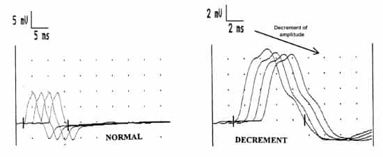
Figure 12 study. Four CMAPs are shown in each tracing. Note that the amplitudes of the responses are the same in a normal muscle, but that a decremental response is recorded in neuromuscular transmission defects.
Please continue to information on EMG/NCV-2 next page
65